The Expanding Role of Deep Learning in Magnetic Resonance Imaging (MRI)
If you believe that medical imaging and deep learning are solely about segmentation, this article is here to prove you wrong. The intersection of these two fields is rich and multifaceted, particularly in the realm of Magnetic Resonance Imaging (MRI). In this article, we will explore various applications of deep neural networks in MRI, demonstrating how deep learning enhances the entire imaging workflow, from signal processing to image reconstruction and beyond.
Understanding the Motivation
The motivation for integrating deep learning into MRI is straightforward yet crucial. Many diagnostic tasks require initial searches to identify abnormalities, quantify measurements, and track changes over time. Moreover, deep learning methods are increasingly being adopted to improve clinical practices. In MRI, deep learning applications can be broadly categorized into two parts:
- Signal Processing Chain: This includes aspects closely related to the physics of MRI, such as image reconstruction, restoration, and registration.
- Post-Reconstruction Applications: This encompasses the use of deep learning on MR images, including segmentation, super-resolution, and medical image synthesis.
To provide a comprehensive overview, we will delve into each of these categories, highlighting key advancements and applications.
Medical Image Reconstruction in MRI
What is Medical Image Reconstruction?
The process of generating MR images can be summarized in a few steps:
- The MRI machine emits a radio frequency (RF) pulse at a specific frequency.
- RF coils transmit the pulse to the targeted area of the body.
- Protons absorb the RF pulse, altering their alignment with the primary magnetic field.
- Once the RF pulse is turned off, the protons relax back to their original alignment, emitting radio waves in the process.
- The spatial information is encoded as measured data during acquisition in the frequency domain, known as k-space.
To visualize this, imagine a 3D volume viewed from above the patient. The k-space contains equivalent information to an MR 2D slice, making it a crucial component of MRI.
Medical Image Reconstruction with Deep Learning
One of the pioneering works in applying deep learning to MRI reconstruction was conducted by Schlemper et al. in 2017. They proposed a framework for reconstructing dynamic sequences of 2D cardiac MR images from under-sampled acquisition data using a deep cascade of convolutional neural networks (CNNs). This approach aimed to accelerate the MRI acquisition process and demonstrated superior performance over traditional methods in terms of reconstruction error and speed while preserving anatomical structures.
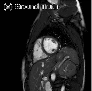
The fastMRI Project: Accelerating MR Imaging with AI
In a collaborative effort, Facebook AI Research (FAIR) and NYU Langone Health launched the fastMRI project, aiming to leverage AI to speed up MRI scans by up to ten times. They introduced the fastMRI dataset, which includes 8,344 volumes and over 1.57 million slices of processed MR images. This dataset serves as a foundation for machine learning breakthroughs in accelerated MR image reconstruction.
The architecture proposed in their recent publication employs a series of U-Net-based models to reconstruct under-sampled k-space data, demonstrating significant advancements in both speed and accuracy.
Medical Image Denoising and Synthesis
Image Generation and Denoising
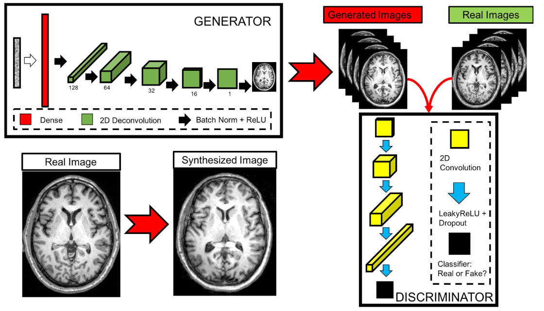
Image synthesis, or generation, involves learning the distribution of data to produce new, realistic images. This technique can be applied to medical images for tasks such as image denoising and translation. Bermudez et al. (2018) explored this by using deep learning to extract quantitative information from acquired images, improving traditional image processing techniques.
They employed a Generative Adversarial Network (GAN) to produce high-quality T1-weighted brain MRI images from a limited dataset, showcasing the potential of deep learning in enhancing image quality.
The Radiologist’s Perspective on Synthetic Images
Despite the comparable quality of synthetic images, expert radiologists noted that certain anatomical abnormalities were more pronounced in these images, highlighting challenges in achieving anatomical accuracy and signal quality. This feedback underscores the importance of continuous refinement in image synthesis techniques.
Medical Image Translation Using Cycle-GAN
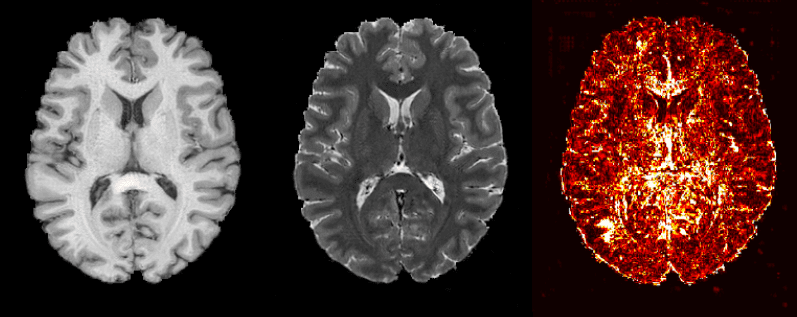
Another innovative application of deep learning in MRI is medical image translation. Welander et al. (2018) utilized Cycle-GAN to translate between T1 and T2 MRI images. By training two generators to learn the forward and inverse mappings, the model achieved more realistic translations, demonstrating the versatility of GANs in medical imaging.
Super-Resolution in Medical Images
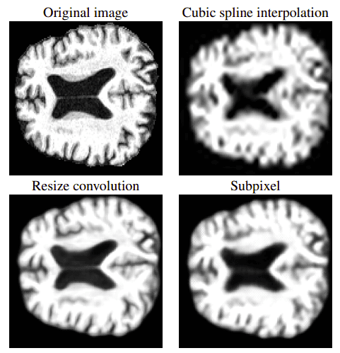
Super-resolution aims to estimate high-resolution images from low-resolution counterparts. Liu et al. (2018) proposed a multi-scale approach that effectively recovers detailed information from low-resolution MRI images. Their model demonstrated significant performance improvements, showcasing the potential of deep learning in enhancing image quality.
Medical Image Registration
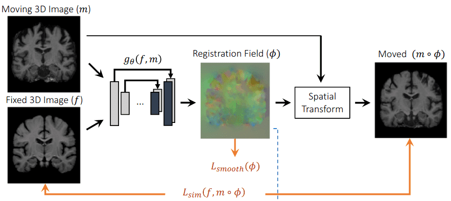
Image registration is the process of aligning different medical images to facilitate meaningful comparisons. This is particularly important for tracking disease progression and treatment planning. Traditional registration methods can be time-consuming, often taking several minutes. However, deep learning approaches like VoxelMorph have emerged, significantly reducing registration time to mere seconds while improving accuracy.
Conclusion
In this article, we provided a broad overview of the applications of deep learning in MRI, emphasizing that its role extends far beyond segmentation. Deep learning enhances various aspects of the MRI workflow, from reconstruction and denoising to image synthesis and registration. While these advancements are promising, challenges remain, particularly in ensuring the generalization of models to real-world clinical settings.
As we move forward, interdisciplinary collaboration among biomedical engineers, deep learning specialists, and radiologists will be crucial in harnessing the full potential of these technologies. For those interested in diving deeper into this field, we recommend exploring the comprehensive work by Lundervold et al. and enrolling in AI for Medicine courses to gain practical insights.
References
- Lundervold, A. S., & Lundervold, A. (2019). An overview of deep learning in medical imaging focusing on MRI. Zeitschrift für Medizinische Physik, 29(2), 102-127.
- Schlemper, J., et al. (2017). A deep cascade of convolutional neural networks for dynamic MR image reconstruction. IEEE Transactions on Medical Imaging, 37(2), 491-503.
- Bermudez, C., et al. (2018). Learning implicit brain MRI manifolds with deep learning. In Medical Imaging 2018: Image Processing (Vol. 10574, p. 105741L).
- Liu, C., et al. (2018). Fusing multi-scale information in convolution network for MR image super-resolution reconstruction. Biomedical Engineering Online, 17(1), 114.
- Sriram, A., et al. (2020). End-to-End Variational Networks for Accelerated MRI Reconstruction. arXiv preprint arXiv:2004.06688.
- Yi, X., et al. (2019). Generative adversarial network in medical imaging: A review. Medical Image Analysis, 58, 101552.
- Cirillo, M. D., et al. (2020). Vox2Vox: 3D-GAN for Brain Tumour Segmentation. arXiv preprint arXiv:2003.13653.
- Welander, P., et al. (2018). Generative adversarial networks for image-to-image translation on multi-contrast MR images-A comparison of CycleGAN and UNIT. arXiv preprint arXiv:1806.07777.
- Sánchez, I., & Vilaplana, V. (2018). Brain MRI super-resolution using 3D generative adversarial networks. arXiv preprint arXiv:1812.11440.
- Balakrishnan, G., et al. (2019). Voxelmorph: a learning framework for deformable medical image registration. IEEE Transactions on Medical Imaging, 38(8), 1788-1800.
- Klein, S., et al. (2009). Elastix: a toolbox for intensity-based medical image registration. IEEE Transactions on Medical Imaging, 29(1), 196-205.
By exploring these advancements, we can appreciate the transformative potential of deep learning in medical imaging, paving the way for improved diagnostic capabilities and patient outcomes.
